What is a neuroforaminal stenosis?
Neuroforaminal stenosis is a narrowing (stenosis) of the nerve exit canals (neuroforamina) between the vertebral bodies of the spine.
The spinal canal contains the spinal cord and the nerve root that originates from it. In this nerve root are motor, autonomic and sensitive nerve fibers. The nerve root initially runs in the spinal canal. It leaves the spinal canal through an exit orifice (neuroforamen) and reaches its final destination: muscles, skin, joints, bladder and other organs.
The exit orifice in the lumbar spine is about 18-22 millimeters high and 5-8 millimeters wide. In the area of the cervical spine (C-spine) 10-14 millimeters, the opening of the neuroforamina is high and 5-7 millimeters wide. When narrowing occurs in the area of the openings, neuroforaminal stenosis occurs - a narrowing of the nerve exit, often called neuroforaminal narrowing.

How do narrowings of the neuroforamina develop?
Such narrowings of the neuroforamina are predominantly caused by bony attachments as part of the degeneration of the spinal column. In the case of advanced signs of wear, calcium deposits and arthrosis of the vertebral arch joints also narrow the root exit openings. The narrowing is also intensified by a loss of height of the intervertebral discs and the associated changes in position. The constriction irritates or even damages the nerves of the spinal cord. Neuroforaminal stenosis is often accompanied by narrowing of the spinal canal (spinal stenosis).
Causes of narrowing of the root exit opening (neuroforaminal stenosis)
A loss of height of the intervertebral disc and a resulting smaller distance between the vertebrae above and below the disc, but also malpositions, can cause a narrowing of the nerve exit opening. Likewise, vertebral slippage, wear and tear, calcification and arthrosis of the small vertebral joints are often responsible for a narrowing of the root exit canal, the narrowing of the neuroforamina.
Symptoms of neuroforaminal stenosis
The symptoms of neuroforaminal stenosis can vary greatly. They range from radiating pain to signs of paralysis in the affected supply area.
In detail: back pain is not the main symptom in isolated neuroforaminal stenosis, but pain radiating to the arms or legs - even bilaterally. The pain in this condition occurs most often with exertion. Therefore, with neuroforaminal stenosis, symptoms usually occur when running or walking.
Relief is felt when bending, while stretching intensifies the symptoms. Patients therefore prefer to remain seated. In addition, the tingling, numbness and sensory disturbances increase. Leg cramps, gait unsteadiness, paralysis of the arms or legs may also occur. Bladder and rectum disorders and potency disorders may occur.
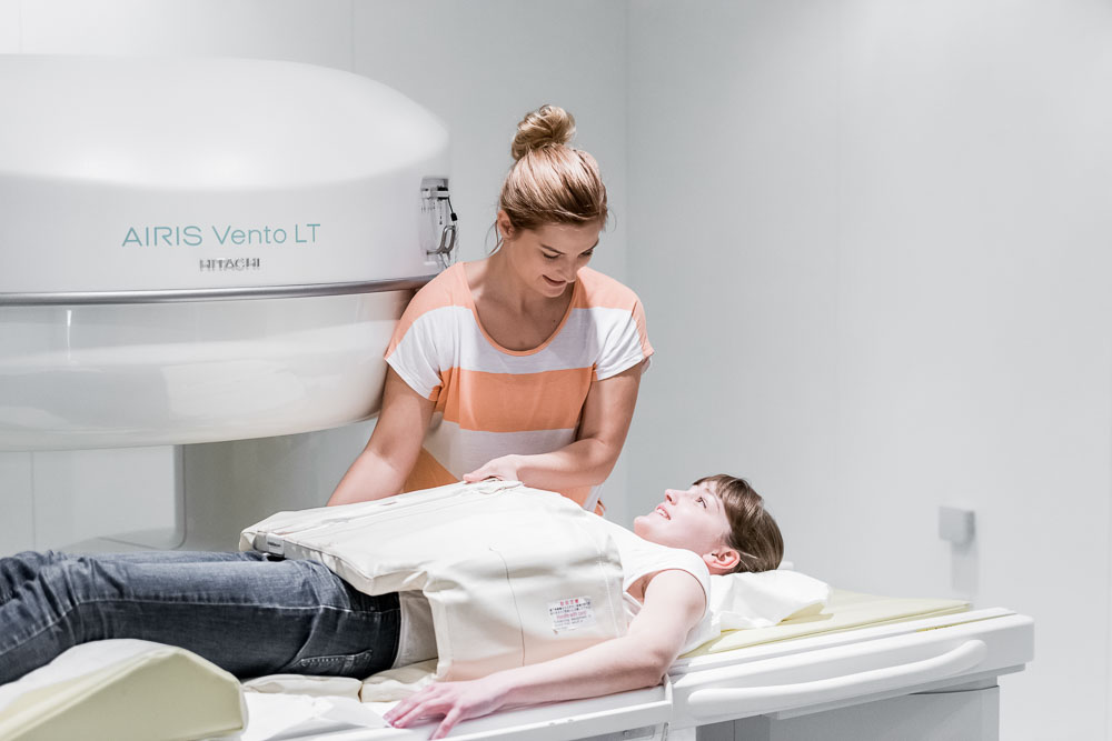
Diagnostics - X-rays, MRI and CT are used
Before treatment, a detailed medical history (in-depth patient interview), a physical and neurological examination, and additional radiological examinations are necessary. Furthermore, an extensive examination with imaging procedures such as X-ray, MRI and CT follows. This is the only way to determine the cause of the complaints.
In order to recommend a sensible, promising therapy for neuroforaminal stenosis to the patient, a very individualized assessment is necessary. To make this possible, doctors are increasingly acting in an interdisciplinary team. These are specialists from a wide variety of fields: orthopedists, radiologists, psychologists, neurologists and, of course, neurosurgeons.
Therapies and treatment of neuroforaminal narrowing.
After careful diagnostics, we decide which therapy and treatment to use for neuroforaminal narrowing in each individual case. We create an individual treatment plan that coordinates the therapy measures. This includes injections, physiotherapy and pain therapy. However, in some cases it is advisable to avoid conservative therapies if they are not feasible due to individual circumstances or if there is a worsening of the stenosis.
Surgery to widen the exit opening of the nerve root.
If conservative spinal stenosis therapies, such as injections of painkillers directly to the affected nerves or physiotherapy, do not have sufficient effect, then microsurgery is suitable. The same is the case when there are insensations, reduction of strength and bladder emptying disorders. Microsurgery provides causal therapy of the stenosis by relieving the nerves by ablating the calcifications and thickened structures. In this way, we widen the exit opening of the nerve root.
Since neuroforaminal stenosis is often combined with spinal stenosis, the spinal canal is also widened during the procedure. Widening of the spinal canal is considered to be the most effective treatment method with the fewest side effects. It is also possible to mill out the nerve root exit holes with the help of a small skin incision in order to achieve optimal widening of the neuroforamina again.
Recovery and recuperation after surgery
Basically, thanks to modern techniques, complaints are alleviated causally and life with all its movements is made easier. Normally, one can expect a convalescence or recovery of a few weeks after surgery. Of course, the length of recovery depends on several factors: These include age and the general health of the patient's body. In some cases, such as when instability is present, stabilization/stiffening surgery may also help as a last option.
Prognosis with neuroforaminal stenosis
You do not have to be afraid of the future despite neuroforaminal stenosis! However, you are challenged to do something for your pain-free or low-pain future.
One of the so-called magic remedies is exercise. Therefore, rehab for neuroforaminal stenosis includes numerous exercises of movement. Cycling is considered ideal because it gives your nerves more space again.
Stay active in sports, because the abdominal and back muscles support your spine. Untrained muscles, on the other hand, lead to tension and new back pain that has nothing to do with the stenosis at all. Always make sure you perform lifting movements correctly or when doing athletic exercises. This will protect your joints and spine. Your body will thank you for it: not only can the risk of neuforaminal stenosis or spinal canal stenosis be alleviated in this way, but a herniated disc or other diseases of the spine can also be reduced.

Information about the article
The article was last checked and updated on November, 16th, 2022.
About the author
Dr. med. Munther Sabarini, MD, is the director and founder of the Avicenna Clinic. As a specialist neurosurgeon, he particularly has expertise in the treatment of spinal disorders. Dr. Munther Sabarini has more than 30 years of professional experience. During this time he treated more than 30,000 patients.
Avicenna Clinic Content Quality Guidelines
All texts and content are written by medically trained, experienced experts in the field. Learn more about our content quality guidelines.
Sources and further literature
F. Schröter, K.-D. Thomann, V. Grosser: Orthopädisch-unfallchirurgische Begutachtung: Handbuch der klinischen Begutachtung. Elsevier Health Sciences, 2019.
A. Tschugg, B. Meyer, M. Stoffel, P. Vajkoczy, F. Ringel, S.-O. Eicker, V. Rhode, C. Thomé: Operative Versorgung der degenerativen Halswirbelsäule. Der Nervenarzt, 06/2018, Springer Medizin, 2018.
The Avicenna Clinic in Berlin is always willing to help you
Since the year 2001, the Avicenna Clinic is based in Berlin. Our doctors have at least 25 years of international experience in their respective fields (neurosurgery, spinal surgery, anesthesia, and orthopedics).
If you have severe back pain, a herniated disc or a suspected herniation, please contact us using the following information: