What are spinal cysts?
Cysts are cavities that form in a tissue and are surrounded by a membrane or wall. A cyst may consist of one or more chambers. The interior is filled with tissue fluid, cerebrospinal fluid, blood, or a mushy content. Cysts can develop anywhere in the body from a variety of causes. Most cysts are harmless.
However, depending on their cause, which organ they affect, how large they are, where they occur and how they behave, the structures can cause problems. For example, when a cyst in the spinal canal of the spine presses on surrounding structures and nerves. This results in the typical symptomatology of so-called nerve root compression syndrome. Like a herniated disc, a cyst is an extra piece of tissue in the spinal canal for which there is no room around the spinal cord. In the spinal canal, it is the nerve roots in particular that can be squeezed by a cyst. Spinal cysts can thus become diseases of the spine, which are manifested by various complaints.
Types of cysts in the spine
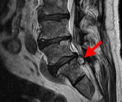
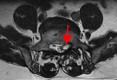
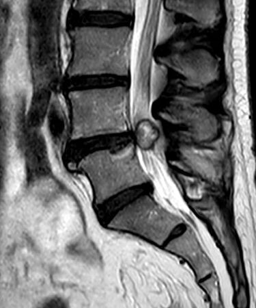
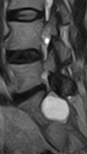
There are two main types of cysts in the spine, the so-called root pocket cysts and synovial cysts (articular cysts, ganglion cysts, pseudocysts). In the vast majority of cases, these are articular cysts, also called articular ganglions. Often, other degenerative changes such as wear, spinal stenosis or spondylolisthesis are also present at the same level as the ganglion cyst.
Cysts have a long history, i.e. past: they were first recognized and described in 1885 - by the British surgeon William Morrant Baker (1839-1896). In 1950, a facet joint cyst was described for the first time as the cause of nerve root compression.
How common are cysts in the spinal canal?
Cysts are rare compared to a narrow lumbar spinal canal (spinal stenosis) or herniated discs (disc herniations). However, they are being diagnosed with increasing frequency thanks to refined imaging using magnetic resonance imaging (MRI). It is therefore not uncommon for them to be an incidental finding.
Studies estimate that 1 percent of synovial cysts are detectable in patients' MRI scans. Synovial cysts can occur in all three spinal segments, i.e., the cervical spine, thoracic spine and lumbar spine, as well as the sacrum. However, they are predominantly found in the lumbar spine. This is where 90 percent of them occur. Of these, the 4th/5th lumbar vertebrae account for 70 percent.
Those affected are predominantly between 58 and 67 years of age, and the diagnosis affects slightly more women than men.
Why do cysts develop?
Cysts can be congenital and acquired. Congenital cysts are formed as a result of defects in the development of embryonic tissue. Acquired cyst develops against the background of inflammatory and degenerative diseases of the spine, as well as injuries and strains. Otherwise, there are many question marks around the formation of joint cysts until today. The formation of joint cysts is therefore not completely clarified. Science assumes that a worn vertebral joint favors a cell proliferation of its joint capsule, which then bulges balloon-like - i.e. cyst-like - into the spinal canal and presses on nerve roots. Thus, degeneration comes to the fore: overloading and inflammation of the small joints can cause the joint capsule to rupture, resulting in the development of a cyst in the spinal canal. Syringomyelia is a cystic formation in the central canal of the spinal cord.
Symptoms - radiating persistent pain
The symptomatology of spinal cysts is closely related to the exact type of cyst:
Ganglion cysts can spread in all directions in the spinal canal. The outwardly directed synovial cysts are usually asymptomatic. It is quite different with inward-facing cysts that grow into the spinal canal. Starting at a size of a few millimeters, joint cysts produce symptoms similar to those of a herniated disc. However, in contrast to classic spinal stenosis, most patients do not complain of back pain, but almost exclusively of radiating, excruciatingly persistent nerve pain, preferably into the leg - depending on which nerve root is pressed and compressed. In fact, the discomfort manifests itself not only when moving, but also when resting. In some cases, symptoms such as numbness or even muscle paralysis also appear. Particularly in older, partially calcified cysts, the symptoms begin insidiously and gradually increase in severity. Only rarely does the symptoms improve, so that surgery is unavoidable in most patients.
Diagnosis of a cysts - anamnesis is always at first
The diagnosis always starts with the first step - and you have to take it: That is the appointment with the doctor! Of course, there are pains that come and go and do not need to be taken seriously. Most of the time, the person affected has a sense for himself/herself whether it is necessary to consult a doctor for this reason. However, there are predominantly indications that clearly signal: Towards the doctor.
The second step is then the anamnesis interview. You explain to the doctor the reasons for your visit and describe your complaints. Normally, he then starts the diagnosis with instrumental methods - after that he has to decide which technique will help him.
When looking for a possible cyst, the doctor will not order an X-ray because it will not effectively help him. This is because the X-ray beam cannot detect a cyst. Therefore, one of the most important methods for diagnosing cysts in the spine is magnetic resonance imaging. It is the standard of imaging, best shows the changes in the spine (narrow spinal canal, herniated disc, cysts, tumors, bleeding, inflammation) and accurately provides the location and extent of joint cysts. If you are living with a pacemaker, the doctor will opt for a computed tomography (CT) scan because an MRI is not feasible for pacemaker patients.
Treatment - how can the problem with the cyst be solved?
Whether and how a cyst needs to be treated is decided by the doctor on a case-by-case basis. Ideally, of course, together with you. Ultimately, the question is: Should only the symptoms be alleviated, or should the problem be solved? To alleviate the symptoms, the doctor may give you a targeted cortisone injection. After that, you may be symptom-free for a few weeks, but that does not solve the cause. In individual cases, there may be spontaneous shrinkage of a synovial cyst, but this is extremely rare. In addition, an emptied cyst fills up with fluid again relatively quickly. As soon as sufficient fluid is again present in the cyst, it presses on the surrounding structures, so that the symptoms are quickly present again. A cyst can only be removed surgically.
The means of choice for definitive treatment is therefore microsurgical decompression surgery. It leads to rapid reduction or complete relief of symptoms. During the minimally invasive operation, a three-centimeter-long skin incision is made in the middle of the back. Under the surgical microscope, the joint cyst is completely detached from the surrounding nerve roots, as adhesions are often found. This is followed by removal of the cyst.
In the case of some cysts caused by parasites, targeted antibiotic therapy often helps.
Prognosis - once the cyst is removed, you will finally be free of pain
Having a cyst is not like having a disease. Only when the cyst expands, make spaces within our bodies, causes pain and other health problems, it becomes a burden for the sufferer. But it is also a warning signal. The important thing is to take this signal seriously. So: go to a doctor (ideally neurosurgeon or orthopedist), when movements and even rest circumstances become an agony. If the joint cyst has been successfully surgically removed from your body, and cannot longer cause you pain, the prognosis for your everyday life is: PAINFREE!

The Avicenna Clinic in Berlin is always willing to help you
Since the year 2001, the Avicenna Clinic is based in Berlin. Our doctors have at least 25 years of international experience in their respective fields (neurosurgery, spinal surgery, anesthesia, and orthopedics).
If you have severe back pain, a herniated disc or a suspected herniation, please contact us using the following information: